Fig. 1
From: The neuropathological diagnosis of Alzheimer’s disease
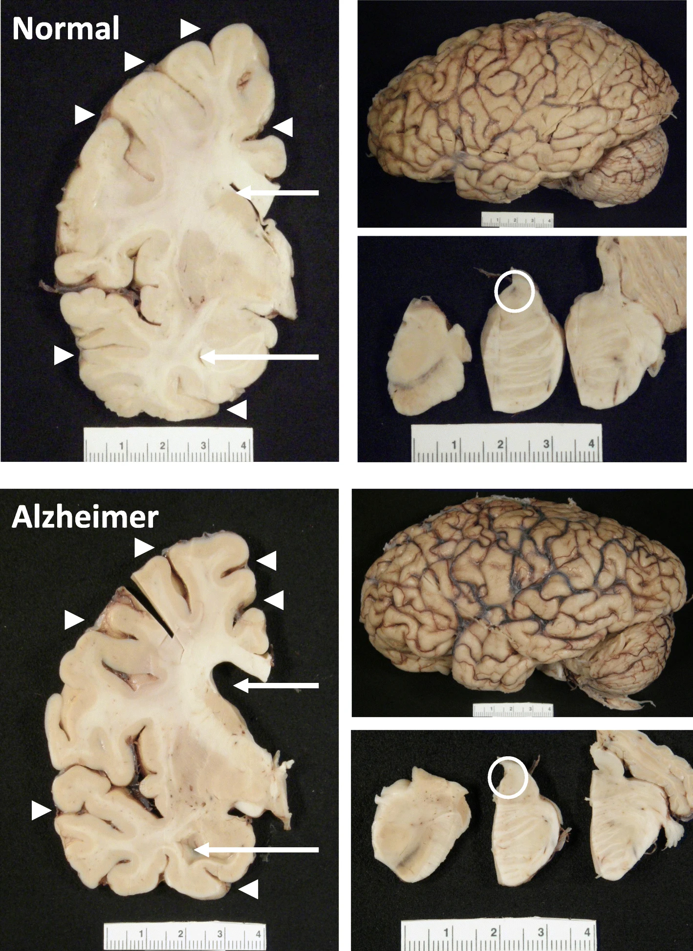
Gross Anatomy of Alzheimer’s Brain. Lateral view of an Alzheimer’s brain can show widening of sulcal spaces and narrowing of gyri compared to a normal brain. This may be more readily observed in coronal sections as indicated by the arrowheads, and this atrophy is often accompanied by enlargement of the frontal and temporal horns of the lateral ventricles as highlighted by the arrows. Additionally, loss of pigmented neurons in the locus coeruleus is commonly observed in the pontine tegmentum as shown with the open circle. None of these features is exclusive to Alzheimer’s disease